

You reach for the morning paper and realize you need to tilt the page toward the kitchen pendant light just to read the stock market numbers.
That subtle daily friction is not just ordinary aging; it is the physiological reality of natural lens yellowing and reduced pupil dilation in low light.
Most people assume fading clarity is an inevitable decline requiring nothing more than stronger drugstore readers, but practicing optometrists prioritize a far more rigorous preventive routine behind closed doors.
⚠️ COMMON MISTAKE
The 60+ Reality Check
Standard eye exams check visual acuity for 20/20 clarity, but they frequently miss contrast sensitivity, macular pigment density, and peripheral integrity—the true markers of functional longevity after 60.
The Early Mechanism: Why the Eye Ages Differently After 60

Cellular aging inside the eye accelerates after your sixth decade.
Cumulative oxidative stress and decades of blue-spectrum light exposure steadily degrade the retinal pigment epithelium, which acts as the metabolic engine supporting your photoreceptors.
At the same time, the gel-like vitreous humor inside your eye begins to liquefy and separate from the retina, making structural monitoring critical to catch micro-tears before symptoms arise.
Capillary density in the choroid layer also decreases with age, reducing nutrient delivery to the back of the eye and making targeted metabolic support essential for preserving sharp central vision.
Cellular Shift Metrics After Age 60
Key physiological changes inside the aging eye
Transitioning from Routine Snellen Charts to Comprehensive Dilation
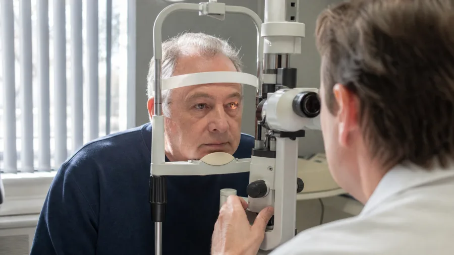
A quick reading chart test at the pharmacy kiosk misses up to 50 percent of asymptomatic eye pathology.
Early-stage glaucoma and peripheral retinal thinning do not hurt, and they do not affect your central reading vision until they are dangerously advanced.
A dilated fundus examination is non-negotiable because the drops allow your eye doctor to directly visualize blood vessel health and optic nerve cupping.
Leading ophthalmological bodies recommend comprehensive dilated exams every one to two years for adults over 60, or annually if you manage systemic conditions like diabetes or hypertension.
Eye Exam Comparison — At a Glance
⏱️ Duration
45 to 60 minutes
📅 Frequency
Every 1 to 2 years
🔍 Detection
Catches glaucoma & tears early
⚠️ Skip If
Never skip dilation after 60
The Dilation Advantage
Dilation opens the pupil wide enough to inspect 80 percent of the retina that remains hidden during quick routine screenings.
Optometric Terms Explained
The Micronutrient Matrix: Targeting Macular Pigment Density

What you put on your plate directly dictates the pigment density protecting your macula.
Specific carotenoids like lutein and zeaxanthin act as internal sunglasses, filtering high-energy blue light before it ever reaches vulnerable photoreceptor cells.
Clinical consensus from major trials like AREDS2 emphasizes that antioxidant vitamins and minerals form the foundation of long-term cellular defense.
You must pair dark leafy greens with healthy fats like olive oil or avocado to unlock their maximum absorption potential in your digestive tract.
💡 PRO TIP
The Fat-Soluble Rule
Lutein is fat-soluble. Eating raw spinach without a healthy fat dressing means your body absorbs only a fraction of the protective pigments your eyes require.
Macular Protection Hierarchy
From daily dietary foundations to targeted support
Environmental and UV Defense Beyond the Summer Months

Ultraviolet radiation does not take a winter vacation.
Cumulative UV-A and UV-B exposure accelerates the clumping of lens proteins, driving cataract formation over decades of outdoor exposure.
You need 100 percent UV-blocking sunglasses even on overcast days and during winter months when glare bounces aggressively off pavement and snow.
Indoor environmental factors matter just as much, requiring you to mitigate dry forced-air heating and adjust digital screen lighting to prevent chronic surface evaporation.
🕶️ Year-Round Ocular Shielding Schedule
Spring Pollen Defense
Flush eyes with saline wash daily to remove airborne allergens before they cause inflammation.
Summer UV400 Wrap
Wear polarized wrap-around sunglasses outdoors to block peripheral scattered rays.
Autumn Wind Mitigation
Use lubricating preservative-free drops when walking in dry autumn breezes.
Winter Glare Control
Put on UV-blocking lenses even on cloudy days to counter snow and pavement reflection.
Environmental Impact Factors
Sources of daily ocular stress after 60
Monitoring Ocular-Systemic Crosslinks: The Vascular Connection

Your eyes serve as a direct, non-invasive window into your entire cardiovascular system.
The microscopic blood vessels inside your retina mirror the health and elasticity of the vessels supplying your heart and brain.
Uncontrolled blood pressure and systemic inflammation directly damage these delicate retinal capillaries, leading to vessel occlusions and ischemic stress.
Proactive management of blood sugar, blood pressure, and systemic hydration is treated by eye doctors as an indispensable pillar of long-term vision preservation.
Vascular Health Approach
Reactive Management
- Waiting for noticeable vision blurring before visiting a clinic
- Ignoring mild morning blood pressure spikes
- Treating eye health completely separate from heart health
- Skipping annual primary care metabolic blood panels
Proactive Ocular Care
- Tracking retinal vessel health during dilated exams
- Maintaining strict daily blood pressure logs
- Hydrating consistently to support systemic microcirculation
- Partnering your optometrist and primary physician together
Vascular Integration Impact
Where Does Your Vision Routine Stand?

Evaluating your current eye care habits requires an honest look at your daily routines and exam history.
Take a moment to audit your last clinical visit: did it include a full pupil dilation, or was it just a quick prescription check for reading glasses?
Check your kitchen pantry for dark leafy greens and your entryway for certified UV400 sunglasses.
Identifying these gaps right now gives you a clear roadmap to discuss with your eye care practitioner at your next scheduled appointment.
Immediate Action Focus
Frequently Asked Questions
Can lifestyle changes actually reverse existing cataracts or macular degeneration?
Lifestyle interventions and targeted nutrition cannot reverse structural tissue damage or dissolve established cataracts. However, they play a powerful role in halting progression, reducing oxidative stress, and preserving remaining functional vision.
How do I know if my dry eyes are age-related or caused by environmental factors?
Age-related dry eye often stems from meibomian gland dysfunction where oil secretions thicken. Environmental dryness worsens symptoms through rapid evaporation; an optometrist can perform specialized meibomian gland expression tests to confirm the exact root cause.
Are over-the-counter blue light blocking glasses necessary for computer use after 60?
Clinical consensus indicates that digital eye strain after 60 is primarily caused by decreased blink rates, uncorrected focusing fatigue, and screen glare rather than blue light emissions from electronic displays.
Is it normal for my vision to fluctuate slightly from morning to evening?
Minor visual fluctuations throughout the day are common and usually relate to tear film stability and mild diurnal intraocular pressure shifts. Consistent or sudden blurring, however, warrants a prompt professional evaluation.
What is the difference between an optometrist and an ophthalmologist when building my care team?
Optometrists provide primary comprehensive eye care, vision testing, and disease screening. Ophthalmologists are medical and surgical doctors who specialize in complex eye diseases, advanced interventions, and surgical procedures like cataract removal.
Securing Your Visual Independence
Preserving your eyesight after 60 requires moving away from passive observation and embracing a structured, daily maintenance routine.
Combine professional clinical partnerships, targeted nutrient intake, and year-round environmental defense to protect your long-term independence.
Take the first step today by booking that long-overdue comprehensive dilated eye exam with your local optometrist.

Meet Edward Green, the founder of GolfingGoal and a seasoned expert in golf. With years of experience on the course and a passion for the sport, Edward has a wealth of knowledge to share. As our Lead Writer, he contributes articles, tutorials, and resources to help readers to improve their skills and reach their full potential on the golf course. Join Our Team!